Strontium »
PDB 1cs7-2grb »
1tjm »
Strontium in PDB 1tjm: Crystallographic Identification of SR2+ Coordination Site in Synaptotagmin I C2B Domain
Protein crystallography data
The structure of Crystallographic Identification of SR2+ Coordination Site in Synaptotagmin I C2B Domain, PDB code: 1tjm
was solved by
Y.Cheng,
S.M.Sequeira,
L.Malinina,
V.Tereshko,
T.H.Sollner,
D.J.Patel,
with X-Ray Crystallography technique. A brief refinement statistics is given in the table below:
| Resolution Low / High (Å) | 20.00 / 1.18 |
| Space group | P 32 2 1 |
| Cell size a, b, c (Å), α, β, γ (°) | 54.644, 54.644, 103.317, 90.00, 90.00, 120.00 |
| R / Rfree (%) | 15.8 / 17.4 |
Strontium Binding Sites:
The binding sites of Strontium atom in the Crystallographic Identification of SR2+ Coordination Site in Synaptotagmin I C2B Domain
(pdb code 1tjm). This binding sites where shown within
5.0 Angstroms radius around Strontium atom.
In total only one binding site of Strontium was determined in the Crystallographic Identification of SR2+ Coordination Site in Synaptotagmin I C2B Domain, PDB code: 1tjm:
In total only one binding site of Strontium was determined in the Crystallographic Identification of SR2+ Coordination Site in Synaptotagmin I C2B Domain, PDB code: 1tjm:
Strontium binding site 1 out of 1 in 1tjm
Go back to
Strontium binding site 1 out
of 1 in the Crystallographic Identification of SR2+ Coordination Site in Synaptotagmin I C2B Domain
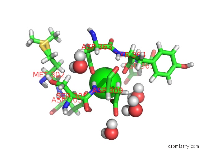
Mono view
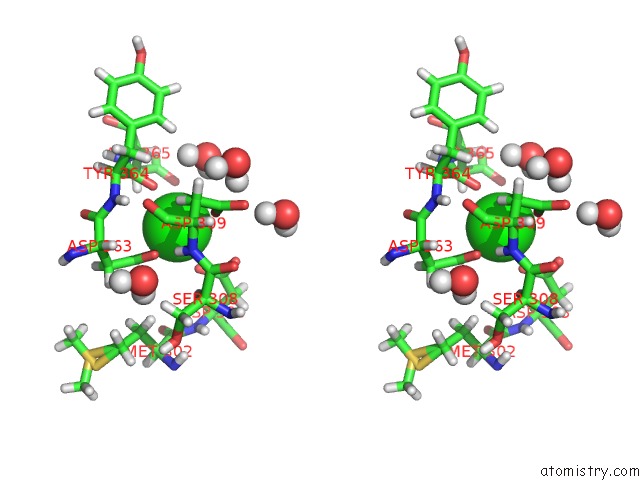
Stereo pair view
Mono view
Stereo pair view
A full contact list of Strontium with other atoms in the Sr binding
site number 1 of Crystallographic Identification of SR2+ Coordination Site in Synaptotagmin I C2B Domain within 5.0Å range:
|
Reference:
Y.Cheng,
S.M.Sequeira,
L.Malinina,
V.Tereshko,
T.H.Sollner,
D.J.Patel.
Crystallographic Identification of CA2+ and SR2+ Coordination Sites in Synaptotagmin I C2B Domain Protein Sci. V. 13 2665 2004.
ISSN: ISSN 0961-8368
PubMed: 15340165
DOI: 10.1110/PS.04832604
Page generated: Thu Oct 10 14:06:09 2024
ISSN: ISSN 0961-8368
PubMed: 15340165
DOI: 10.1110/PS.04832604
Last articles
F in 7MR5F in 7MON
F in 7MOG
F in 7MOO
F in 7MML
F in 7MMI
F in 7MMK
F in 7MMJ
F in 7MMG
F in 7MMF