Strontium »
PDB 3ilg-4rne »
4rby »
Strontium in PDB 4rby: First X-Ray Structure of Rna Containing Guanosine Phosphorodithioate
Protein crystallography data
The structure of First X-Ray Structure of Rna Containing Guanosine Phosphorodithioate, PDB code: 4rby
was solved by
P.S.Pallan,
M.Egli,
with X-Ray Crystallography technique. A brief refinement statistics is given in the table below:
| Resolution Low / High (Å) | 50.00 / 1.19 |
| Space group | C 1 2 1 |
| Cell size a, b, c (Å), α, β, γ (°) | 40.932, 35.015, 31.873, 90.00, 128.80, 90.00 |
| R / Rfree (%) | 18.1 / 21.3 |
Strontium Binding Sites:
The binding sites of Strontium atom in the First X-Ray Structure of Rna Containing Guanosine Phosphorodithioate
(pdb code 4rby). This binding sites where shown within
5.0 Angstroms radius around Strontium atom.
In total 2 binding sites of Strontium where determined in the First X-Ray Structure of Rna Containing Guanosine Phosphorodithioate, PDB code: 4rby:
Jump to Strontium binding site number: 1; 2;
In total 2 binding sites of Strontium where determined in the First X-Ray Structure of Rna Containing Guanosine Phosphorodithioate, PDB code: 4rby:
Jump to Strontium binding site number: 1; 2;
Strontium binding site 1 out of 2 in 4rby
Go back to
Strontium binding site 1 out
of 2 in the First X-Ray Structure of Rna Containing Guanosine Phosphorodithioate
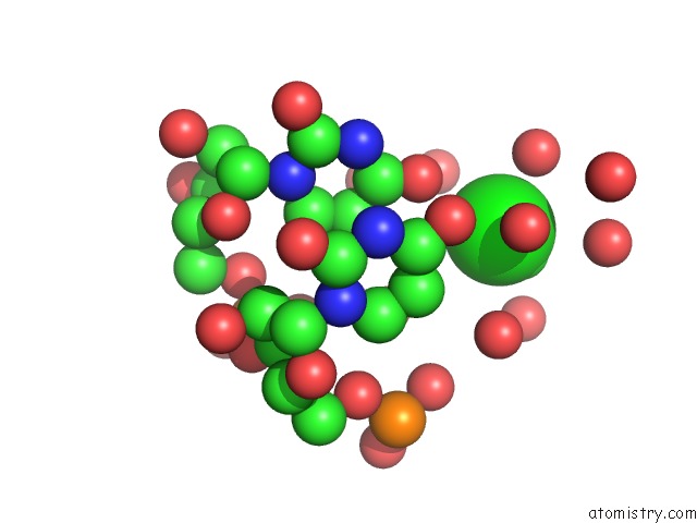
Mono view

Stereo pair view
Mono view
Stereo pair view
A full contact list of Strontium with other atoms in the Sr binding
site number 1 of First X-Ray Structure of Rna Containing Guanosine Phosphorodithioate within 5.0Å range:
|
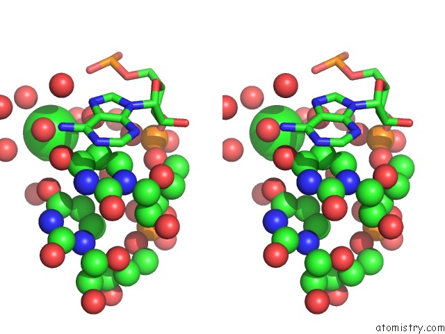
Strontium binding site 2 out of 2 in 4rby
Go back to
Strontium binding site 2 out
of 2 in the First X-Ray Structure of Rna Containing Guanosine Phosphorodithioate
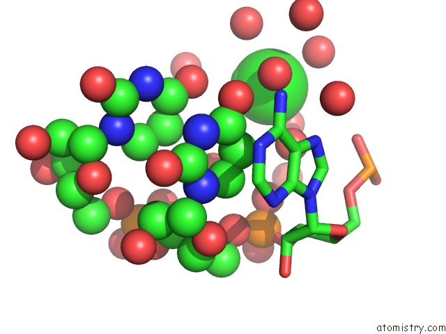
Mono view
Stereo pair view
Mono view
Stereo pair view
A full contact list of Strontium with other atoms in the Sr binding
site number 2 of First X-Ray Structure of Rna Containing Guanosine Phosphorodithioate within 5.0Å range:
|
Reference:
P.S.Pallan,
X.Yang,
M.Sierant,
N.D.Abeydeera,
T.Hassell,
C.Martinez,
M.Janicka,
B.Nawrot,
M.Egli.
Crystal Structure, Stability and Sirna Activity of Phosphorodithioate-Modified Rnas To Be Published.
Page generated: Fri Oct 11 05:46:09 2024
Last articles
Fe in 2YXOFe in 2YRS
Fe in 2YXC
Fe in 2YNM
Fe in 2YVJ
Fe in 2YP1
Fe in 2YU2
Fe in 2YU1
Fe in 2YQB
Fe in 2YOO