Strontium »
PDB 4rum-6ny9 »
6nhh »
Strontium in PDB 6nhh: Rhodobacter Sphaeroides BC1 with Azoxystrobin
Enzymatic activity of Rhodobacter Sphaeroides BC1 with Azoxystrobin
All present enzymatic activity of Rhodobacter Sphaeroides BC1 with Azoxystrobin:
1.10.2.2;
1.10.2.2;
Protein crystallography data
The structure of Rhodobacter Sphaeroides BC1 with Azoxystrobin, PDB code: 6nhh
was solved by
D.Xia,
F.Zhou,
C.A.Yu,
with X-Ray Crystallography technique. A brief refinement statistics is given in the table below:
| Resolution Low / High (Å) | 39.29 / 3.00 |
| Space group | P 1 21 1 |
| Cell size a, b, c (Å), α, β, γ (°) | 89.309, 154.655, 100.939, 90.00, 95.36, 90.00 |
| R / Rfree (%) | 26.4 / 28.2 |
Other elements in 6nhh:
The structure of Rhodobacter Sphaeroides BC1 with Azoxystrobin also contains other interesting chemical elements:
| Iron | (Fe) | 10 atoms |
Strontium Binding Sites:
The binding sites of Strontium atom in the Rhodobacter Sphaeroides BC1 with Azoxystrobin
(pdb code 6nhh). This binding sites where shown within
5.0 Angstroms radius around Strontium atom.
In total 3 binding sites of Strontium where determined in the Rhodobacter Sphaeroides BC1 with Azoxystrobin, PDB code: 6nhh:
Jump to Strontium binding site number: 1; 2; 3;
In total 3 binding sites of Strontium where determined in the Rhodobacter Sphaeroides BC1 with Azoxystrobin, PDB code: 6nhh:
Jump to Strontium binding site number: 1; 2; 3;
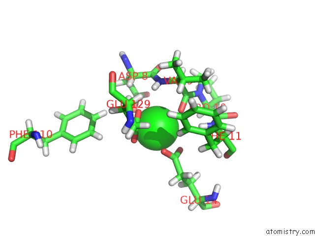
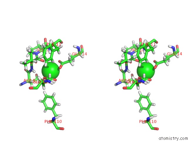
Strontium binding site 1 out of 3 in 6nhh
Go back to
Strontium binding site 1 out
of 3 in the Rhodobacter Sphaeroides BC1 with Azoxystrobin
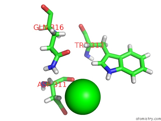
Mono view
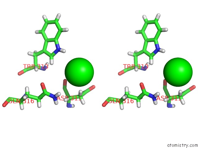
Stereo pair view
Mono view
Stereo pair view
A full contact list of Strontium with other atoms in the Sr binding
site number 1 of Rhodobacter Sphaeroides BC1 with Azoxystrobin within 5.0Å range:
|
Strontium binding site 2 out of 3 in 6nhh
Go back to
Strontium binding site 2 out
of 3 in the Rhodobacter Sphaeroides BC1 with Azoxystrobin
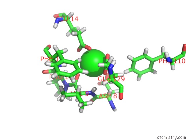
Mono view
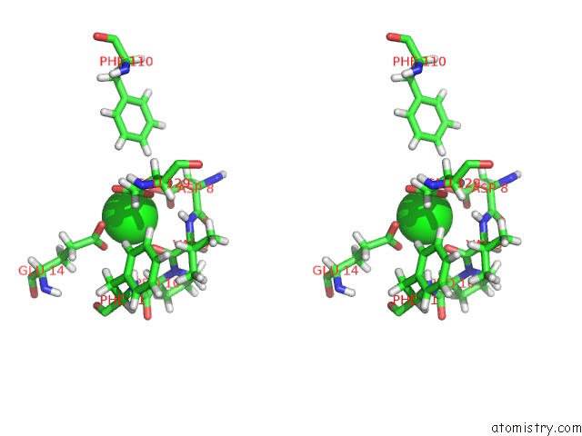
Stereo pair view
Mono view
Stereo pair view
A full contact list of Strontium with other atoms in the Sr binding
site number 2 of Rhodobacter Sphaeroides BC1 with Azoxystrobin within 5.0Å range:
|
Strontium binding site 3 out of 3 in 6nhh
Go back to
Strontium binding site 3 out
of 3 in the Rhodobacter Sphaeroides BC1 with Azoxystrobin
Mono view
Stereo pair view
Mono view
Stereo pair view
A full contact list of Strontium with other atoms in the Sr binding
site number 3 of Rhodobacter Sphaeroides BC1 with Azoxystrobin within 5.0Å range:
|
Reference:
L.Esser,
F.Zhou,
C.A.Yu,
D.Xia.
Crystal Structure of Bacterial CYTOCHROMEBC1IN Complex with Azoxystrobin Reveals A Conformational Switch of the Rieske Iron-Sulfur Protein Subunit. J.Biol.Chem. V. 294 12007 2019.
ISSN: ESSN 1083-351X
PubMed: 31182483
DOI: 10.1074/JBC.RA119.008381
Page generated: Fri Oct 11 06:45:34 2024
ISSN: ESSN 1083-351X
PubMed: 31182483
DOI: 10.1074/JBC.RA119.008381
Last articles
Zn in 9MJ5Zn in 9HNW
Zn in 9G0L
Zn in 9FNE
Zn in 9DZN
Zn in 9E0I
Zn in 9D32
Zn in 9DAK
Zn in 8ZXC
Zn in 8ZUF