Strontium »
PDB 8cvd-8oyr »
8cvh »
Strontium in PDB 8cvh: Structure of L289F Hyoscyamine 6-Beta Hydroxylase in Complex with Vanadyl, Succinate, and 6-Oh-Hyoscyamine
Enzymatic activity of Structure of L289F Hyoscyamine 6-Beta Hydroxylase in Complex with Vanadyl, Succinate, and 6-Oh-Hyoscyamine
All present enzymatic activity of Structure of L289F Hyoscyamine 6-Beta Hydroxylase in Complex with Vanadyl, Succinate, and 6-Oh-Hyoscyamine:
1.14.11.11;
1.14.11.11;
Protein crystallography data
The structure of Structure of L289F Hyoscyamine 6-Beta Hydroxylase in Complex with Vanadyl, Succinate, and 6-Oh-Hyoscyamine, PDB code: 8cvh
was solved by
E.W.Wenger,
A.K.Boal,
J.M.Bollinger,
C.Krebs,
with X-Ray Crystallography technique. A brief refinement statistics is given in the table below:
| Resolution Low / High (Å) | 43.75 / 2.03 |
| Space group | P 1 21 1 |
| Cell size a, b, c (Å), α, β, γ (°) | 44.915, 79.102, 56.276, 90, 111.25, 90 |
| R / Rfree (%) | 20.1 / 25 |
Other elements in 8cvh:
The structure of Structure of L289F Hyoscyamine 6-Beta Hydroxylase in Complex with Vanadyl, Succinate, and 6-Oh-Hyoscyamine also contains other interesting chemical elements:
| Vanadium | (V) | 1 atom |
Strontium Binding Sites:
The binding sites of Strontium atom in the Structure of L289F Hyoscyamine 6-Beta Hydroxylase in Complex with Vanadyl, Succinate, and 6-Oh-Hyoscyamine
(pdb code 8cvh). This binding sites where shown within
5.0 Angstroms radius around Strontium atom.
In total 2 binding sites of Strontium where determined in the Structure of L289F Hyoscyamine 6-Beta Hydroxylase in Complex with Vanadyl, Succinate, and 6-Oh-Hyoscyamine, PDB code: 8cvh:
Jump to Strontium binding site number: 1; 2;
In total 2 binding sites of Strontium where determined in the Structure of L289F Hyoscyamine 6-Beta Hydroxylase in Complex with Vanadyl, Succinate, and 6-Oh-Hyoscyamine, PDB code: 8cvh:
Jump to Strontium binding site number: 1; 2;
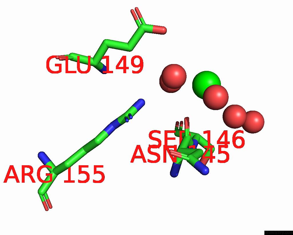
Strontium binding site 1 out of 2 in 8cvh
Go back to
Strontium binding site 1 out
of 2 in the Structure of L289F Hyoscyamine 6-Beta Hydroxylase in Complex with Vanadyl, Succinate, and 6-Oh-Hyoscyamine

Mono view

Stereo pair view
Mono view
Stereo pair view
A full contact list of Strontium with other atoms in the Sr binding
site number 1 of Structure of L289F Hyoscyamine 6-Beta Hydroxylase in Complex with Vanadyl, Succinate, and 6-Oh-Hyoscyamine within 5.0Å range:
|
Strontium binding site 2 out of 2 in 8cvh
Go back to
Strontium binding site 2 out
of 2 in the Structure of L289F Hyoscyamine 6-Beta Hydroxylase in Complex with Vanadyl, Succinate, and 6-Oh-Hyoscyamine
Mono view

Stereo pair view
Mono view
Stereo pair view
A full contact list of Strontium with other atoms in the Sr binding
site number 2 of Structure of L289F Hyoscyamine 6-Beta Hydroxylase in Complex with Vanadyl, Succinate, and 6-Oh-Hyoscyamine within 5.0Å range:
|
Reference:
E.S.Wenger,
A.K.Boal,
J.M.Bollinger,
C.Krebs.
Structure of the L289F H6H Cyclization Ferryl-Mimicking Complex To Be Published.
Page generated: Fri Oct 11 07:48:05 2024
Last articles
Cl in 5ZFBCl in 5ZFA
Cl in 5ZF9
Cl in 5ZDR
Cl in 5ZF8
Cl in 5ZDQ
Cl in 5ZF7
Cl in 5ZF4
Cl in 5ZEQ
Cl in 5ZDL