Strontium »
PDB 4rum-6ny9 »
6nhh »
Strontium in PDB 6nhh: Rhodobacter Sphaeroides BC1 with Azoxystrobin
Enzymatic activity of Rhodobacter Sphaeroides BC1 with Azoxystrobin
All present enzymatic activity of Rhodobacter Sphaeroides BC1 with Azoxystrobin:
1.10.2.2;
1.10.2.2;
Protein crystallography data
The structure of Rhodobacter Sphaeroides BC1 with Azoxystrobin, PDB code: 6nhh
was solved by
D.Xia,
F.Zhou,
C.A.Yu,
with X-Ray Crystallography technique. A brief refinement statistics is given in the table below:
| Resolution Low / High (Å) | 39.29 / 3.00 |
| Space group | P 1 21 1 |
| Cell size a, b, c (Å), α, β, γ (°) | 89.309, 154.655, 100.939, 90.00, 95.36, 90.00 |
| R / Rfree (%) | 26.4 / 28.2 |
Other elements in 6nhh:
The structure of Rhodobacter Sphaeroides BC1 with Azoxystrobin also contains other interesting chemical elements:
| Iron | (Fe) | 10 atoms |
Strontium Binding Sites:
The binding sites of Strontium atom in the Rhodobacter Sphaeroides BC1 with Azoxystrobin
(pdb code 6nhh). This binding sites where shown within
5.0 Angstroms radius around Strontium atom.
In total 3 binding sites of Strontium where determined in the Rhodobacter Sphaeroides BC1 with Azoxystrobin, PDB code: 6nhh:
Jump to Strontium binding site number: 1; 2; 3;
In total 3 binding sites of Strontium where determined in the Rhodobacter Sphaeroides BC1 with Azoxystrobin, PDB code: 6nhh:
Jump to Strontium binding site number: 1; 2; 3;
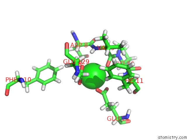
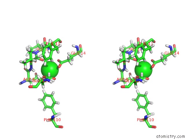
Strontium binding site 1 out of 3 in 6nhh
Go back to
Strontium binding site 1 out
of 3 in the Rhodobacter Sphaeroides BC1 with Azoxystrobin
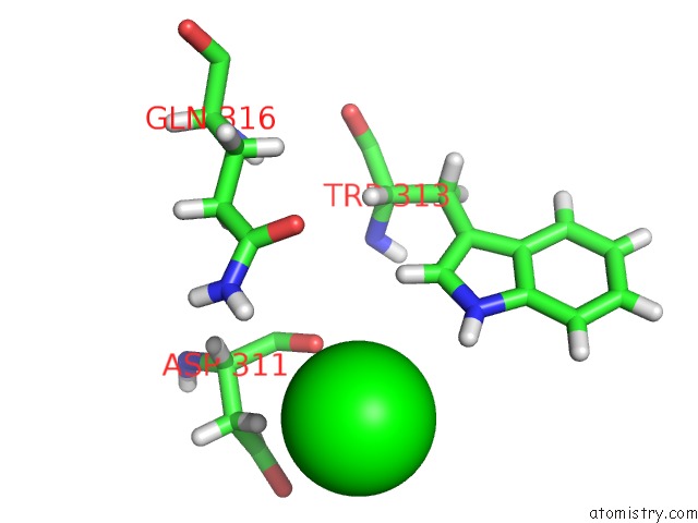
Mono view
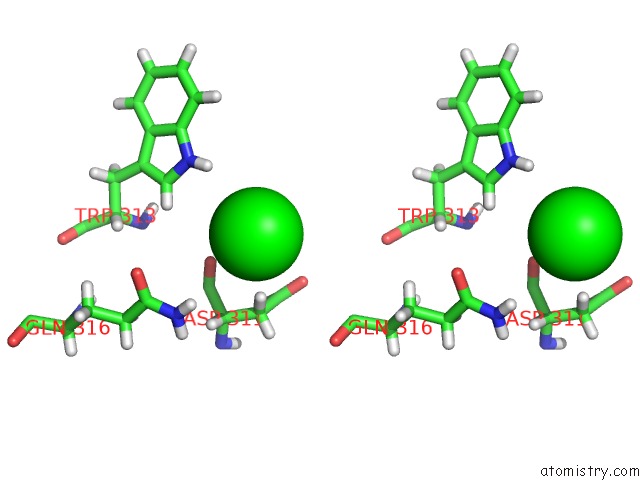
Stereo pair view
Mono view
Stereo pair view
A full contact list of Strontium with other atoms in the Sr binding
site number 1 of Rhodobacter Sphaeroides BC1 with Azoxystrobin within 5.0Å range:
|
Strontium binding site 2 out of 3 in 6nhh
Go back to
Strontium binding site 2 out
of 3 in the Rhodobacter Sphaeroides BC1 with Azoxystrobin
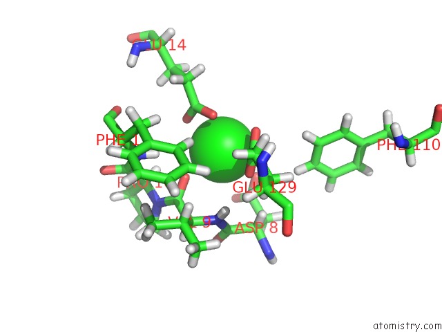
Mono view
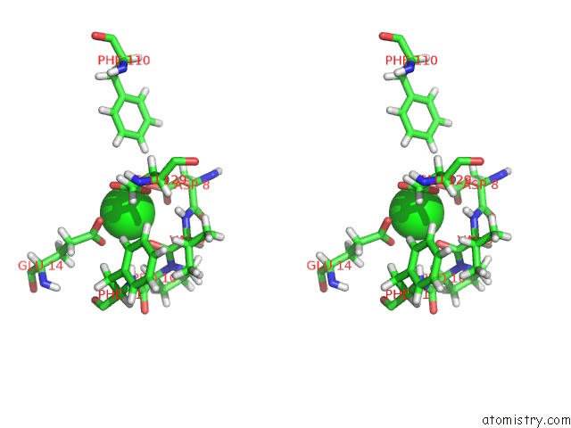
Stereo pair view
Mono view
Stereo pair view
A full contact list of Strontium with other atoms in the Sr binding
site number 2 of Rhodobacter Sphaeroides BC1 with Azoxystrobin within 5.0Å range:
|
Strontium binding site 3 out of 3 in 6nhh
Go back to
Strontium binding site 3 out
of 3 in the Rhodobacter Sphaeroides BC1 with Azoxystrobin
Mono view
Stereo pair view
Mono view
Stereo pair view
A full contact list of Strontium with other atoms in the Sr binding
site number 3 of Rhodobacter Sphaeroides BC1 with Azoxystrobin within 5.0Å range:
|
Reference:
L.Esser,
F.Zhou,
C.A.Yu,
D.Xia.
Crystal Structure of Bacterial CYTOCHROMEBC1IN Complex with Azoxystrobin Reveals A Conformational Switch of the Rieske Iron-Sulfur Protein Subunit. J.Biol.Chem. V. 294 12007 2019.
ISSN: ESSN 1083-351X
PubMed: 31182483
DOI: 10.1074/JBC.RA119.008381
Page generated: Fri Oct 11 06:45:34 2024
ISSN: ESSN 1083-351X
PubMed: 31182483
DOI: 10.1074/JBC.RA119.008381
Last articles
I in 8KEUI in 8K4Z
I in 8K0T
I in 8JLN
I in 8J9S
I in 8JLJ
I in 8IKP
I in 8IKS
I in 8ING
I in 8IKB